inSiliScope
A deterministic in-silico microscope. inSiliScope generates an endless synthetic sample and images it the way a microscope would: as a single-molecule localisation (SMLM) movie or as a widefield image, with a physical PSF and camera noise model. Two samples can be mounted: a field of cells (cell body, nucleus, cytoplasm, microtubules, nuclear pores, fluorophore sites; the microtubules and the pores labelled in two colours) and DNA-origami nanorulers (rods with three spots of docking strands 80 nm apart).
The live microscope in your browser; the Quickstart shows what to try.
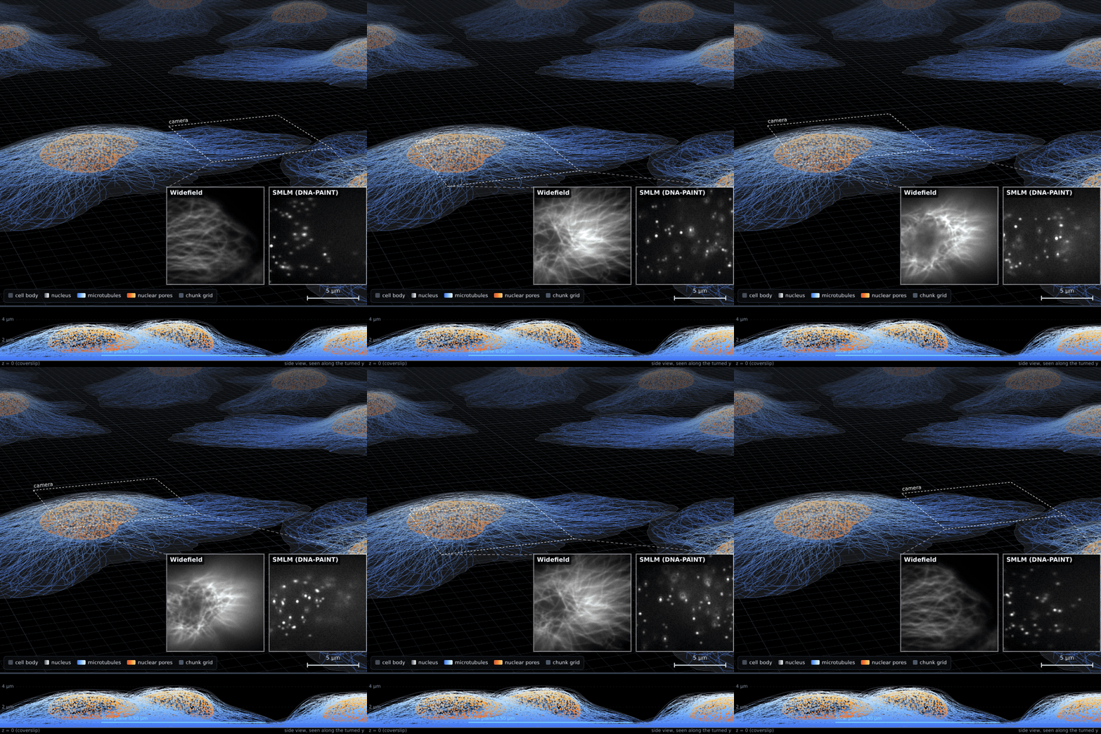
The cell field in the viewer, tilted and in perspective, and what its live microscope sees in the dashed camera box. More in the Gallery.
One C++ world model, three consumers:
| Consumer | What it is | Where |
|---|---|---|
| Viewer | Pan/zoom/tilt/turn through the field in your browser, a live microscope (bleaching where you look, the Micro-Manager config presets) that records TIFF stacks (WASM); setups saved and loaded as files in mixable parts (instrument, settings, sample, measurement); cell animations exported as MP4/WebM/GIF | Open the viewer, how to use it |
| Micro-Manager adapter | A hub (mmgr_dal_inSiliScope.dll) with one device per part of the microscope (camera, stages, objective, filters, lasers, lamp, sample): move the stage, change focus, run live acquisitions, sequences and z stacks against a sample that never changes |
Micro-Manager |
| webSMLM block | The same core as a single generated JS file for webSMLM | Quickstart |
Why it exists
Testing localisation and analysis software needs data where the truth is known, and testing acquisition software needs a sample that behaves like a sample: the same cell is in the same place every time the stage returns to it, bleaching persists, focus matters. inSiliScope provides both from one source of truth.
Key properties
- Address-based determinism. Every cell, microtubule and dye is a pure function of
(seed, address). Any window of the infinite field can be generated alone, in any order, on any platform, with identical results. See World model. - Bit-exact across targets. The RNG and the geometry are the same bit for bit natively and in WASM, guarded by golden vectors in CI.
- Dyes and a light path. Each structure has its own label: a dye of the library (FPbase spectra; AF647, mEos3.2, ATTO 655, mEGFP, ...) in a mode: dSTORM, PALM (with the pre-converted state), DNA-PAINT (with the free imager's background) or WideField (every dye at once, bleaching by its photon budget). Lasers, dichroic, emission filter and the camera's QE curve decide what is excited and detected, and at which wavelength (the PSF follows). See Dyes and light path.
- Two modalities. Fluorescence with a diffraction PSF (a scalar Gibson-Lanni model with Zernike aberration modes, double helix) or a Gaussian. BrightField (exploration): transmitted light through the cells' refractive index, partially coherent multislice wave optics, a speed/precision quality setting.
- Realistic camera. sCMOS and EMCCD noise chains with per-pixel maps, background and illumination profiles.
- The light's geometry. One laser spot in the objective's back focal plane (epi, HILO, TIRF) or several coherent beams interfering to a 2D, 3D or lattice SIM pattern that steps with the frame (Structured illumination).
- More than one sample. The geometry of a sample is separate from the photophysics and the imaging: the cell
field and the nanorulers share the dyes, the light path, the PSF and the camera.
Switch in the viewer's Sample select, Micro-Manager's
SampleHolderor the cli's--specimen; see Samples. Fiducial beads can be added to either, for drift correction; they can be imaged alone too (the bead sample), and so can a fluorescent slide, a uniform dye layer. - Built to extend. Samples, targets, photophysics and modalities are separate layers; see Extending.
Reading guide
- New here: the Quickstart (a tour of the viewer), then the Gallery.
- Want the models: Samples (the cell field's world model and structures, the nanorulers) and Physics (dyes and light path, photophysics, optics, camera, validation).
- Want to add something: Extending and the Roadmap.
- Timings: Benchmarks.
Status
Pre-1.0. The numbers are reproducible for a given version and seed (world_version is recorded in outputs once
introduced; see the roadmap). The Direct3D 11 widefield path is checked on software and Wine adapters; reports from
real GPUs are welcome.
License
The source is BSD-3-Clause. The distributed Micro-Manager DLL is GPL-3.0; the viewer and the webSMLM block are
BSD-3-Clause. See the repository's LICENSE and
THIRD_PARTY_NOTICES.md.